 Our Live Cams


| Size: 367.37 MB[/center]
Wolf Parkinsons White Syndrome : A Case Based Discussion
Published 5/2024
MP4 | Video: h264, 1920x1080 | Audio: AAC, 44.1 KHz
Language: English
| Duration: 0h 38m
Etiology , Causes, Clinical Features, Pathophysiology Complications with Management of Wolf Parkinsons White Syndrome
What you'll learn
What is Wolf Parkinsons White Syndrome WPW
What are clinical Features of WPW Syndrome
What are ECG Findings of WPW Syndrome
Complications and Management of WPW Syndrome
Requirements
Basic Medical Knowledge
Description
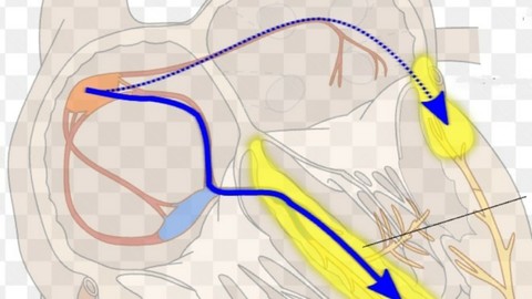
Etiology , Causes, Clinical Features, Pathophysiology Complications with Management of Wolf Parkinsons White Syndrome.Wolff Parkinson White Syndrome (WPW) is considered to be a congenital abnormality that involves the presence of abnormal electrical conductive circuits between the atria and ventricles. The disorder includes accessory electrical pathways that bypass the AV node. This activity reviews the evaluation and treatment of WPW by an interprofessional healthcare team.Objectives:Identify the etiology of Wolff Parkinson White syndrome.Outline the evaluation of Wolff Parkinson White syndrome.Review the management options available for Wolff Parkinson White syndrome.Describe the interprofessional team strategies for improving care coordination and improve patient outcomes in patients with Wolff Parkinson White syndrome.Wolff-Parkinson-White (WPW) syndrome is a congenital cardiac preexcitation syndrome that arises from abnormal cardiac electrical conduction through an accessory pathway that can result in symptomatic and life-threatening arrhythmias. The hallmark electrocardiographic (ECG) finding of WPW pattern or preexcitation consists of a short PR interval and prolonged QRS with an initial slurring upstroke ("delta" wave) in the presence of sinus rhythm. The term WPW syndrome is reserved for an ECG pattern consistent with the above-described findings along with the coexistence of a tachyarrhythmia and clinical symptoms of tachycardia such as palpitations, episodic lightheadedness, presyncope, syncope, or even cardiac arrest.The normal heart consists of two electrically insulated units, the atria and the ventricles. These units are connected by a conduction system that allows for normal cardiac synchrony and function. The cardiac electrical potential originates from the sinoatrial node of the right atrium and propagates through the atria to the atrioventricular (AV) node. The action potential is delayed in the AV node and is then quickly transmitted through the His-Purkinje system to the ventricular myocytes allowing for rapid ventricular depolarization and synchronized contraction.[1] Patients with WPW syndrome have an accessory pathway that violates the electrical isolation of the atria and ventricles, which can allow electrical impulses to bypass the AV node. In some settings, this pathway can result in the transmission of abnormal electrical impulses leading to malignant tachyarrhythmias. The ECG findings of the WPW pattern are caused by the fusion of ventricular preexcitation through the accessory pathway and normal electrical conduction. Most patients with WPW pattern will never develop arrhythmia and will remain asymptomatic. Some accessory pathways will not manifest the described typical ECG findings, and as a result, some patients can develop a tachyarrhythmia with no prior ECG evidence that the pathway exists. These are referred to as concealed bypass tracts.EtiologyWPW pattern arises from the fusion of ventricular preexcitation through the accessory pathway and normal electrical conduction through the AV node. This accessory pathway is thought to arise from chamber myocardium during improper early atrial and ventricular folding in cardiac embryogenesis. As a result, electrically conductive myocardial bundles violate the normal electrical insulation of the atrium and ventricle, forming the accessory pathway.[1][7] This pathway usually has non-decremental or non-delayed conduction, which is in contrast to the properties of the normal AV node. The electrical conducting characteristics of the accessory pathway can vary and depend upon factors such as the speed of conduction, direction of conduction, and refractory period. These characteristics, along with location and number of pathways, will determine how the pathway may be involved in the initiation or transmission of an arrhythmia leading to WPW syndrome.
Overview
Section 1: Introduction
Lecture 1 Introduction
Section 2: Lecture 2 : WPW Syndrome : Etiology , Pathophysiology and Discovery
Lecture 2 Lecture 2 : WPW Syndrome : Etiology , Pathophysiology and Discovery
Section 3: Lecture 3 : Pathophysiology
Lecture 3 Lecture 3 : Pathophysiology
Section 4: Lecture 4 : Clinical Features of WPW Syndrome
Lecture 4 Lecture 4 : Clinical Features of WPW Syndrome
Section 5: Lecture 5 : Evaluation of WPW Syndrome
Lecture 5 Lecture 5 : Evaluation of WPW Syndrome
Section 6: Lecture 6 : Management of WPW Syndrome
Lecture 6 Lecture 6 : Management of WPW Syndrome
Course intended for Allied health professionals and health professionals
https://fikper.com/HPyIB4lTPM/Wolf.Parkinsons.White.Syndrome.A.case.Based.Discussion.rar.html
https://rapidgator.net/file/bf46ce905d08e49ddd8fb4a3847eb1a4/Wolf.Parkinsons.White.Syndrome.A.case.Based.Discussion.rar
https://katfile.com/60blqvluqd15/Wolf.Parkinsons.White.Syndrome.A.case.Based.Discussion.rar
Free search engine download: Wolf Parkinsons White Syndrome A case Based Discussion


 Reply With Quote